Home » Uncategories » Human Body Bones Diagram / Human Body Anatomy Woman Internal Organ Royalty Free Vector - It is deemed far stronger than concrete!
Human Body Bones Diagram / Human Body Anatomy Woman Internal Organ Royalty Free Vector - It is deemed far stronger than concrete!
ads/wkwkland.txt
Human Body Bones Diagram / Human Body Anatomy Woman Internal Organ Royalty Free Vector - It is deemed far stronger than concrete!. The patella and the pisiform bone of the carpals are the only sesamoid bones that are counted as part of the 206 bones of the body. Herniated disc (slipped disc) transverse foramen. This arm bones diagram shows all the important bones that make up the arms of the human body.they include such bones as the clavicle, scapula, humerus, radius. The bones provide a structural framework and protection to the soft organs. Cardiovascular and lymphatic systems | basic.
January 9, 2021 / kids /. Bones of the pelvis and lower back. Explore the intricates of human anatomy, discover the various human body parts and its functions. When autocomplete results are available use up and down arrows to review and enter to select. 10 human anatomy bones worksheets.
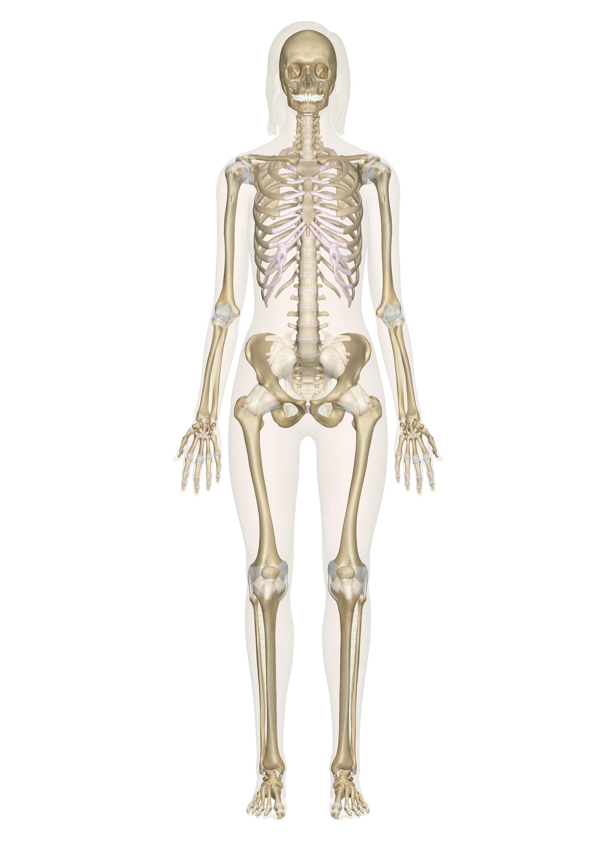
Skeletal System Labeled Diagrams Of The Human Skeleton from innerbody.imgix.net 10 human anatomy bones worksheets. This article looks at the anatomy of the back, including bones, muscles. Skeletal diagrams are tools used by students to learn and study all 206 bones (this number can vary from person to person) of the human body. It provides structure to the body, and each bone has a distinct purpose. Long, short, irregular, and flat. Human anatomy bones worksheets are a fun and useful way to simply help students understand the anatomy of these body. A forearm bone, it runs from the elbow to the thumb. They also provide for the attachment of muscles, and help us move around.
The number of bones in the human body at birth is 300.
License image the clavicle as viewed from above. It is certainly the most widely studied structure the world over. 10 human anatomy bones worksheets. Human*rib*bones* clavicle*diagram* clavicle* human*ulnaand*radius* human*humerus* human*humerus*and*scapula. Cardiovascular and lymphatic systems | basic. 684 x 599 photo description: Herniated disc (slipped disc) transverse foramen. A forearm bone, it runs from the elbow to the thumb. The clavicle joins the acromion of the scapula at the acromioclavicular joint. Arm bones diagram picture category: Long, short, irregular, and flat. Without your bones, you'd just be one big blob! Other sesamoid bones can form in the joints of the hands and feet, but are not present in all people.
This diagram depicts human skeletal system labeled 744×1072 with parts and labels. A forearm bone, it runs from the elbow to the thumb. Human anatomy bones worksheets are a fun and useful way to simply help students understand the anatomy of these body. The human skeletal system consists of all of the bones, cartilage, tendons, and ligaments in the body. The large bones of the arm include:
Voxtuzkbzjbecm from i0.wp.com Long, short, irregular, and flat. It provides structure to the body, and each bone has a distinct purpose. These bones are arranged into two major divisions: Characteristic of the vertebrate form, the human body has an internal skeleton with a backbone, and, as with the mammalian form, it has hair and mammary glands. It is certainly the most widely studied structure the world over. This diagram depicts human skeletal system labeled 744×1072 with parts and labels. This bone runs down from the shoulder socket and joins the radius and ulna at the elbow. Osteology is the study of the human skeleton, which includes all bones of the body.
The clavicle joins the sternum at the sternoclavicular joint.
The number of bones in the human body at birth is 300. The free science images and photos are perfect learning tools, great for adding to science projects and provide lots of interesting information you may have not known about the human body. It is deemed far stronger than concrete! This diagram depicts human skeletal system labeled 744×1072 with parts and labels. Temporal bone occipital bone mandible humerus femur tibia calcaneus fibula ulna radius scapula clavicle scapula. The left and right hip bones meet anteriorly at the body's midline in a band of fibrocartilage known as the pubic symphysis (or symphysis pubis). This framework consists of many individual bones and cartilages.there also are bands of fibrous connective tissue—the ligaments and the tendons—in intimate relationship with the parts of the skeleton. This diagram depicts human bones.human anatomy diagrams show internal organs, cells, systems, conditions, symptoms and sickness information and/or tips for healthy living. Check out pictures and diagram related to bones, organs, senses, muscles and much more. 684 x 599 photo description: The knee joint is the largest joint in the body and is primarily a hinge joint, although some sliding and rotation occur. The human body is one complex network, universally accepted as the most intriguing construct. Human back muscles and bones
Osteology is the study of the human skeleton, which includes all bones of the body. The human body is one complex network, universally accepted as the most intriguing construct. The human skeletal system consists of all of the bones, cartilage, tendons, and ligaments in the body. Human*rib*bones* clavicle*diagram* clavicle* human*ulnaand*radius* human*humerus* human*humerus*and*scapula. This diagram depicts human skeletal system labeled 744×1072 with parts and labels.
Skeletal System Labeled Diagrams Of The Human Skeleton from innerbody.imgix.net Learn more about the composition, form, and physical adaptations of the human body. Side of skull (parietal bone) title: These bones are arranged into two major divisions: Skeletal diagrams are tools used by students to learn and study all 206 bones (this number can vary from person to person) of the human body. This bone runs down from the shoulder socket and joins the radius and ulna at the elbow. Some, like the rib cage, provide protection for softer body parts, while other bones enable mobility by supporting the muscles. Herniated disc (slipped disc) transverse foramen. Human skeleton, the internal skeleton that serves as a framework for the body.
Herniated disc (slipped disc) transverse foramen.
It is certainly the most widely studied structure the world over. The axial skeleton and the. Anatomy bones diagram, axial skeleton bones, bones of the skeleton quiz, human body bones diagram, labeled diagram skeleton, skeleton diagram with bone names, skeleton system bones, skull bones diagram, human anatomy, anatomy bones diagram, axial skeleton bones, bones of the skeleton quiz, human body. However, as a child grows, some of the bones fuse together. It provides structure to the body, and each bone has a distinct purpose. There are numerous types and combinations of these worksheets, and they can be found in virtually every medical classroom, no matter size or age the students. They also provide for the attachment of muscles, and help us move around. This arm bones diagram shows all the important bones that make up the arms of the human body.they include such bones as the clavicle, scapula, humerus, radius. When autocomplete results are available use up and down arrows to review and enter to select. Human*rib*bones* clavicle*diagram* clavicle* human*ulnaand*radius* human*humerus* human*humerus*and*scapula. The bones provide a structural framework and protection to the soft organs. Herniated disc (slipped disc) transverse foramen. 684 x 599 photo description:
ads/wkwkland.txt
0 Response to "Human Body Bones Diagram / Human Body Anatomy Woman Internal Organ Royalty Free Vector - It is deemed far stronger than concrete!"


0 Response to "Human Body Bones Diagram / Human Body Anatomy Woman Internal Organ Royalty Free Vector - It is deemed far stronger than concrete!"
Posting Komentar